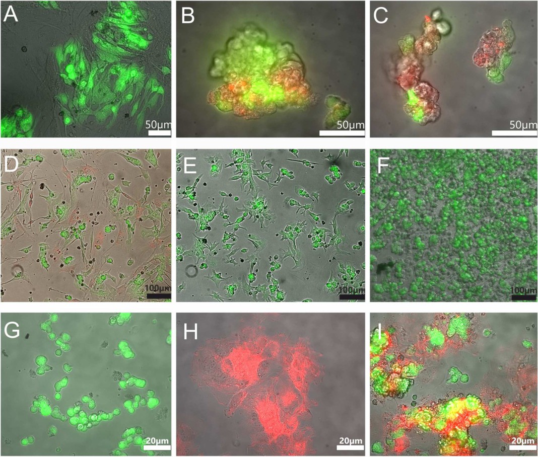
Fig. 1. Generation of in vitro grown micro-tissues for cardiac cell replacement therapy. Micro-tissues were generated on thermo-responsive polymer surfaces. Cell detachment resulted in the formation of aggregates of MSCs and ES-CMs. For low-dose MSC/ES-CM experiments, optimized ratios of MSCs and ES-CMs (green) were plated (A). Micro-tissues formed under low-dose MSC/ES-CM conditions by cold-induced detaching are shown (CMs: green, MSCs: red) (B,C). Micro-tissues that were passed through an injection cannula readily attached to conventional tissue culture surfaces (D). Coating of thermo-responsive polymer surfaces with fibronectin allowed for the formation of micro-tissues from pure ES-CMs without addition of MSCs (E) and could be detached from the surface of the culture dish and re-plated after passing the injection cannula without affecting cell integrity (F). iPS-CMs lifted from thermo-responsive polymers were cultured for 2 hours on tissue culture plastics and did not show signs of adhesion (G). MSCs prepared from the bone marrow of red fluorescent protein (Tomato) expressing mice, cultured for 2 hours on tissue culture plastics, adhered well (H). Micro-tissues composed of iPS-CMs and MSCs plated for 2 hours on tissue culture plastic adhered well and anchored the iPS-CMs to the surface (I). Note that the MSCs were stained by CMTPX (red fluorescence) in images B, C, and D. Cardiomyocytes express eGFP and display green fluorescence (all panels).